- Home
- Diabetes Complications
- Diabetic Foot Ulcer Treatment and Warning Signs
Diabetic Foot Ulcers: Treatment, Warning Signs, Home Care, and Prevention
Diabetic foot ulcers are open wounds that require prompt attention because reduced sensation, poor circulation, and infection can delay healing and increase the risk of serious complications. This article explains the warning signs, evidence-based treatment, safe home care, and practical steps that can help prevent future ulcers.
Written by Dr. Albana Greca, MD, MMedSc, Family Physician
Medically reviewed by Dr. Ruden Cakoni, MD, Endocrinologist
Last reviewed: July 2026
Quick Answer
A diabetic foot ulcer is an open wound below the ankle in a person with diabetes. It often develops because reduced sensation allows pressure or injury to go unnoticed, while poor circulation, infection, foot deformity, and high glucose can make healing more difficult.
Every new foot ulcer needs prompt professional assessment. Treatment usually combines wound cleaning and debridement when appropriate, pressure offloading, infection treatment only when infection is present, circulation assessment, glucose management, suitable dressings, and close follow-up. A hot, red, swollen, black, foul-smelling, rapidly worsening, or deeply infected foot requires urgent care.
A diabetes-related foot ulcer is not simply a skin problem. The wound may involve pressure injury, neuropathy, peripheral artery disease, infection, bone involvement, and foot deformity at the same time. Healing is most successful when these problems are assessed together by a multidisciplinary foot-care team.
Do not wait for severe pain before seeking help. People with diabetic peripheral neuropathy may have little or no pain even when the ulcer is deep or infected.
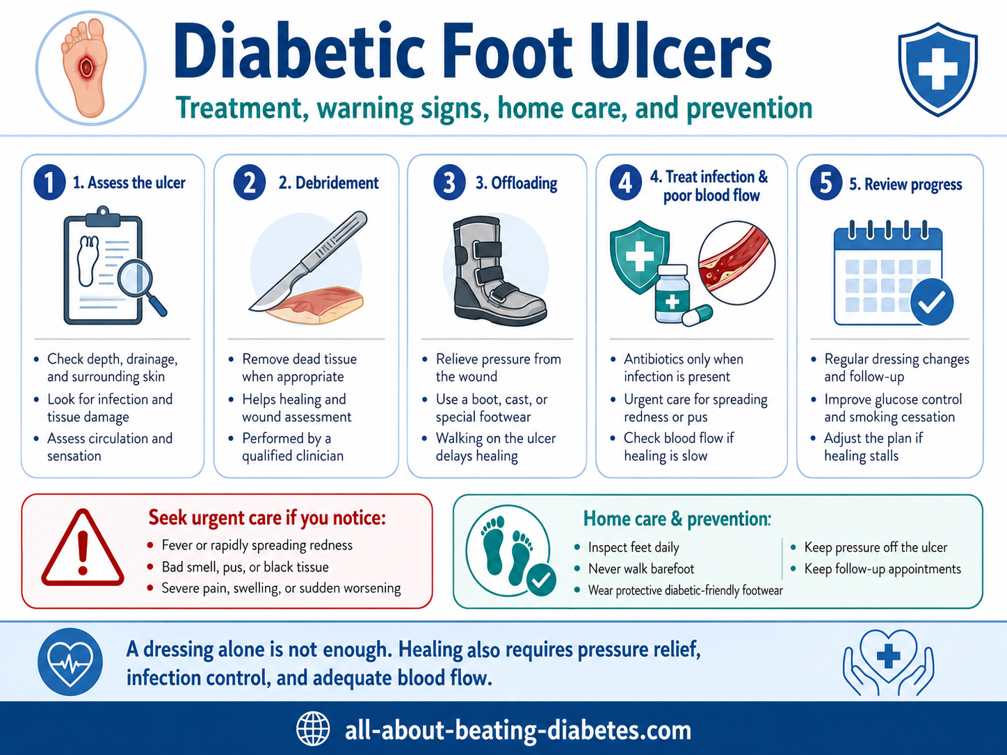

What Is a Diabetic Foot Ulcer?
A diabetes-related foot ulcer is a break in the skin below the ankle that extends through at least part of the dermis. It may be shallow or extend into fat, tendon, joint, or bone. Common locations include the ball of the foot, heel, toes, sides of the foot, and areas under callus or deformity.
Foot ulcers are serious because infection can spread into deeper tissues or bone, and reduced blood flow may prevent healing. Untreated ulcers can lead to hospitalization, gangrene, amputation, sepsis, and loss of mobility.
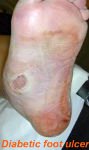
Foot ulcers vary in depth and appearance. A clinician must assess the wound rather than judging severity from a photograph alone.
What Causes Diabetic Foot Ulcers?
Most ulcers result from several interacting factors rather than one cause.
Loss of protective sensation
Neuropathy can reduce the ability to feel pressure, heat, pain, blisters, small stones, tight shoes, or minor injuries. Repeated pressure then damages the skin without warning.
Pressure and foot deformity
Callus, bunions, hammer toes, prominent metatarsal heads, limited joint movement, previous amputation, and Charcot foot can concentrate pressure in a small area. Shoes that are too tight, worn, or poorly fitted can add friction and shear.
Peripheral artery disease
Diabetes increases the risk of narrowed leg arteries. Poor blood flow reduces oxygen and nutrient delivery, slows healing, and increases the risk of tissue loss. Warning signs may include absent pulses, cold feet, color change, pain with walking, rest pain, or wounds that fail to improve—but peripheral artery disease can also be painless.
Infection
Infection usually begins after the skin barrier is broken. It may remain local or spread into deeper tissue, tendon, joint, or bone. High glucose can impair immune function and may make infection more difficult to control.
Additional risk factors
- Previous foot ulcer or lower-limb amputation
- Kidney disease, especially dialysis
- Poor vision or inability to inspect the feet
- Smoking
- Long diabetes duration
- Limited mobility
- Walking barefoot
- Inappropriate nail or callus treatment
- Social, financial, or access barriers that delay care
Symptoms and Warning Signs
A diabetic foot ulcer may appear as an open sore, crack, blister, draining callus, or dark area. Because neuropathy can reduce pain, visual and temperature changes may be more important than discomfort.
| Finding | What It May Mean | Action |
|---|---|---|
| New break in the skin, blister, or draining callus | Early ulcer or pressure injury | Arrange prompt assessment and reduce pressure |
| Increasing redness, warmth, swelling, pus, odor, or pain | Possible infection | Same-day medical assessment |
| Black, blue, pale, or rapidly darkening tissue | Severe ischemia, necrosis, or gangrene | Urgent vascular and surgical assessment |
| Deep wound, exposed tendon or bone, or ulcer that probes to bone | Possible deep infection or osteomyelitis | Urgent specialist evaluation |
| Fever, chills, confusion, rapid breathing, low blood pressure, or severe weakness | Possible spreading infection or sepsis | Emergency care |
| Hot, red, swollen foot with little pain and possible deformity | Possible active Charcot neuroarthropathy | Stop weight-bearing and seek same-day care |
Emergency Warning
Seek same-day urgent care for a new ulcer with spreading redness, warmth, swelling, pus, foul odor, black tissue, exposed bone, sudden deformity, severe pain, or a foot that is much hotter than the other foot.
Call emergency services for confusion, collapse, severe breathing difficulty, rapidly spreading discoloration, sepsis symptoms, or a person who appears critically ill.
How a Diabetic Foot Ulcer Is Assessed
A comprehensive assessment looks beyond the surface of the wound. The clinician may evaluate:
- Ulcer location, size, depth, duration, drainage, callus, and tissue type
- Redness, warmth, swelling, tenderness, pus, odor, and systemic infection signs
- Whether tendon, joint, or bone may be involved
- Foot pulses, capillary refill, temperature, color, and peripheral artery disease risk
- Protective sensation and other neuropathy findings
- Foot shape, pressure points, gait, shoes, and previous ulcers or amputations
- Glucose pattern, HbA1c, kidney function, nutrition, smoking, and medicines
Circulation testing
Testing may include ankle and toe pressures, Doppler waveforms, toe-brachial index, transcutaneous oxygen measurement, or vascular imaging. An ankle-brachial index can be misleading when diabetes has caused stiff, calcified arteries, so several tests may be needed.
When the ulcer is ischemic, fails to improve, or has signs of severe blood-flow limitation, early vascular assessment is essential. Revascularization may be required to restore blood flow before healing is possible.
Testing for infection and bone involvement
Foot infection is diagnosed clinically—not by wound culture alone. An open ulcer is commonly colonized by bacteria, but colonization is not the same as infection.
When infection is suspected, a properly collected tissue specimen is generally more useful than a superficial swab. Tests for suspected osteomyelitis may include a probe-to-bone examination, plain X-ray, inflammatory markers, MRI, and bone sampling when needed.
How Diabetic Foot Ulcers Are Treated
1. Pressure offloading
Pressure relief is one of the most important treatments for a neuropathic plantar ulcer. Walking normally on the wound can repeatedly damage new tissue, even when the ulcer is covered.
For an uncomplicated neuropathic ulcer under the forefoot or midfoot, guidelines recommend a non-removable knee-high offloading device as the first choice when it is safe and tolerated. Options may include a total-contact cast or a removable walker made non-removable by the clinical team.
When a non-removable device is unsuitable, a removable knee-high or ankle-high device may be considered. The patient must use it during all weight-bearing activity. Ordinary shoes or standard “diabetic shoes” are not adequate treatment for many active plantar ulcers.
Infection, ischemia, balance, falls risk, ulcer location, work, home conditions, and the ability to attend follow-up can change the offloading choice. Do not buy or apply a cast or pressure-relief device without professional fitting.
2. Debridement and wound-bed care
Debridement removes callus, dead tissue, debris, or infected material when appropriate. Sharp debridement is often used, but the method and timing depend on blood flow, infection, wound depth, pain, bleeding risk, and clinician expertise.
Do not cut callus, scrape the wound, or remove black tissue at home. Dry stable black tissue on a severely ischemic toe or heel may require a different approach until circulation is assessed.
3. Dressings
The dressing should protect the ulcer, manage drainage, avoid trauma during removal, and support a moist—not waterlogged—healing environment. Options include foams, alginates, hydrofibers, hydrogels, films, and other products.
No single dressing is best for every ulcer. Selection depends on drainage, depth, infection, location, surrounding skin, cost, and how often it can be changed. Antimicrobial dressings are not automatically required.
4. Infection treatment
Antibiotics are used when the ulcer is clinically infected. They should not be prescribed merely because an ulcer is open or because a culture grows bacteria.
Current infection guidelines advise against systemic or topical antibiotics for a clinically uninfected ulcer when the goal is to prevent infection or accelerate healing. Unnecessary antibiotics can cause adverse effects and antimicrobial resistance.
Moderate or severe infection may require hospitalization, intravenous antibiotics, urgent drainage, removal of dead tissue, or surgery. Infection with severe ischemia requires urgent input from both surgical and vascular specialists.
5. Restore blood flow when needed
Peripheral artery disease can prevent healing despite excellent dressings and glucose control. Revascularization may be performed by endovascular procedures or open surgery, depending on the arterial disease, anatomy, health status, and local expertise.
6. Glucose, nutrition, and whole-person care
Glucose should be managed safely because persistent hyperglycemia can impair immune function and healing. However, suddenly pursuing very tight control without considering hypoglycemia, kidney function, appetite, infection, and frailty can be dangerous.
Wound healing also requires adequate calories, protein, vitamins, minerals, and hydration. Routine high-dose vitamin or supplement products are not proven to heal ulcers and may interact with medicines. A dietitian can help when there is weight loss, poor intake, kidney disease, or malnutrition risk.
Smoking cessation, blood-pressure treatment, cholesterol management, kidney care, and cardiovascular risk reduction are also part of limb protection. Our overview of diabetes and foot complications explains how neuropathy and circulation interact.
Advanced Wound Treatments: When Are They Used?
Advanced therapies are considered only after the fundamentals are addressed: debridement, offloading, infection treatment, circulation assessment or revascularization, glucose management, and appropriate dressings.
Hyperbaric oxygen therapy
Hyperbaric oxygen therapy is not routine treatment for every diabetic foot ulcer. Current IWGDF guidance conditionally supports considering it as an adjunct for selected neuro-ischemic or ischemic ulcers when good standard care has failed and an established program is available.
Evidence for improved healing is mixed, and reliable evidence that it prevents amputation is limited. Treatment is time-consuming, costly, and not suitable for everyone.
Topical oxygen
Topical oxygen may be considered as an adjunct for selected nonhealing ulcers after standard care has failed, but evidence quality is limited and devices differ.
Negative-pressure wound therapy
Negative-pressure wound therapy may be useful for selected postsurgical diabetic foot wounds. It is not automatically superior for every nonsurgical ulcer and requires careful protection of vessels, bone, and surrounding skin.
Skin substitutes and placental-derived products
Selected cellular, acellular, or placental-derived products may be considered for ulcers that do not heal with good standard care. Evidence, cost, availability, wound suitability, and insurance coverage vary. These products cannot compensate for untreated infection, continued pressure, or inadequate blood flow.
Surgery
Surgery may be needed to drain infection, remove dead or infected bone, correct a deformity, reduce pressure, cover a wound, or restore blood flow. Amputation may be life-saving when infection or tissue death cannot be controlled, but limb-preserving treatment is considered whenever safe and feasible.
How to Care for a Diabetic Foot Ulcer at Home
Home care should follow a written plan from the wound-care team. It does not replace professional treatment.
Daily home-care checklist
- Wash and dry your hands before and after dressing care.
- Clean the wound only with the solution recommended by the clinical team.
- Use the prescribed dressing and change it at the instructed interval.
- Keep the dressing clean, secure, and protected from water.
- Use the offloading device every time you bear weight unless told otherwise.
- Check the wound and surrounding skin for redness, swelling, odor, drainage, or color change.
- Check glucose according to your diabetes plan and take medicines as prescribed.
- Attend every wound, podiatry, diabetes, and vascular appointment.
- Contact the team promptly if the ulcer becomes larger, deeper, wetter, darker, hotter, or more painful.
What not to do
- Do not soak the foot.
- Do not walk barefoot or in socks alone.
- Do not use hydrogen peroxide, bleach, strong iodine, acids, corn removers, or caustic antiseptics unless specifically prescribed.
- Do not apply herbal pastes, raw garlic, honey, oils, powders, or household remedies to the ulcer.
- Do not cut callus, dead tissue, or nails near the wound yourself.
- Do not massage a swollen or ischemic foot.
- Do not use heating pads, hot-water bottles, or direct heat.
- Do not stop antibiotics early or take leftover antibiotics.
- Do not resume normal walking because the wound “looks better.”
Exercise recommendations must be adapted while the ulcer is active. Ask the foot team about safe non-weight-bearing activity. Walking for circulation is not appropriate when it repeatedly loads an open plantar ulcer.
How Is Healing Monitored?
The wound should be measured and documented regularly. Clinicians review area, depth, tissue quality, drainage, surrounding skin, infection, blood flow, pressure relief, and adherence to the treatment plan.
A wound that is not making meaningful progress should trigger reassessment. Reasons may include unrelieved pressure, unrecognized ischemia, infection, osteomyelitis, poor nutrition, edema, an unsuitable dressing, smoking, or an incorrect diagnosis.
Even after the skin closes, the foot remains at high risk. “Healed” tissue is vulnerable, and recurrence prevention requires protective footwear, gradual return to activity, daily inspection, and regular professional care.
How to Prevent Diabetic Foot Ulcers
- Inspect the tops, soles, heels, and spaces between toes every day.
- Use a mirror or ask another person for help when necessary.
- Wash with lukewarm water and dry carefully, especially between the toes.
- Moisturize dry skin, but not between the toes.
- Wear properly fitted shoes and clean socks indoors and outdoors.
- Check inside shoes for stones, sharp seams, moisture, or foreign objects.
- Do not treat corns, calluses, or ingrown nails with over-the-counter acids or blades.
- Stop smoking and manage glucose, blood pressure, cholesterol, and kidney risk.
- Have a comprehensive professional foot examination at least annually and more often when neuropathy, peripheral artery disease, deformity, previous ulcer, or amputation is present.
Our diabetic foot-care guide provides a practical daily prevention routine.
Can a Diabetic Foot Ulcer Heal Completely?
Many ulcers can close with timely comprehensive treatment. Healing depends on wound depth, blood flow, infection, pressure relief, nutrition, diabetes control, kidney health, smoking, and the ability to follow the treatment plan.
Closure does not mean the underlying risk has disappeared. Neuropathy, deformity, and circulation disease often remain, so recurrence prevention becomes the next stage of treatment.
Doctor’s Note
When I assess a diabetic foot ulcer, I do not ask only which dressing to use. I ask whether the patient can feel the foot, whether the wound is infected, whether blood can reach it, whether pressure is still damaging it, and whether bone is involved. Missing any one of these factors can delay healing or threaten the limb.
Most Asked Questions
Should every diabetic foot ulcer receive antibiotics?
No. Antibiotics are used for clinical infection. An uninfected ulcer should not receive antibiotics simply to prevent infection or promote healing.
Can I walk on a diabetic foot ulcer?
Usually the pressure must be reduced substantially. The correct offloading device depends on ulcer location, infection, blood flow, balance, and other factors. Follow the foot-care team’s weight-bearing instructions.
Can I clean the ulcer with hydrogen peroxide?
Do not use hydrogen peroxide or another strong antiseptic unless the wound clinician specifically instructs you to do so. These products can damage healing tissue.
Is hyperbaric oxygen effective?
It may help selected ischemic or neuro-ischemic ulcers when standard treatment has failed, but evidence is mixed and it is not routine first-line therapy.
How do I know whether the ulcer has reached bone?
You cannot determine this reliably at home. A deep ulcer, exposed bone, a positive probe-to-bone examination, X-ray, MRI, laboratory findings, and sometimes a bone sample may be used.
Why does the ulcer not hurt?
Neuropathy may reduce pain sensation. Lack of pain does not mean the wound is minor or uninfected.
Educational safety note: This page does not diagnose or treat an individual wound. A new or worsening foot ulcer requires professional examination. Do not perform debridement, change antibiotics, or alter weight-bearing without clinical guidance.
Related Questions
- Will Lowering Blood Sugar Reduce Foot Swelling?
- What Causes Diabetic Foot Swelling in the Afternoon?
- What Causes Painful Purple or Blue Diabetic Feet?
- Can Medicine Improve Blood Flow to Diabetic Feet?
Related Resources
- Diabetic Foot Care
- Diabetic Peripheral Neuropathy
- Diabetes and Foot Problems
- Diabetic Foot Symptoms
- Diabetes Complications and Prevention
- Understanding Diabetes Blood Sugar Levels
References
- American Diabetes Association. Retinopathy, Neuropathy, and Foot Care: Standards of Care in Diabetes—2026.
- International Working Group on the Diabetic Foot. Guidelines on Prevention and Management of Diabetes-Related Foot Disease—2023.
- IWGDF/IDSA. Diagnosis and Treatment of Diabetes-Related Foot Infections—2023.
- IWGDF. Offloading Foot Ulcers in Persons With Diabetes—2023.
- IWGDF. Interventions to Enhance Healing of Foot Ulcers—2023.
- NIDDK. Diabetes and Foot Problems.